LATEST NEWS
Surgical Ophthalmic Oncology: A Collaborative Open Access Surgical Textbook
In 2014, the First Eye Cancer Working Day was hosted by The Curie Institute in Paris, France. There, we decided to develop a Collaborative Open Access Surgical Textbook (COAST) aimed at offering guidance and international outreach for ophthalmologists in countries without eye cancer specialists. The resultant 5-year effort was spearheaded by Drs. Sonal Chaugule, Santosh [..] Read More...
International Multicenter Cooperative Study Supported by The Eye Cancer Foundation
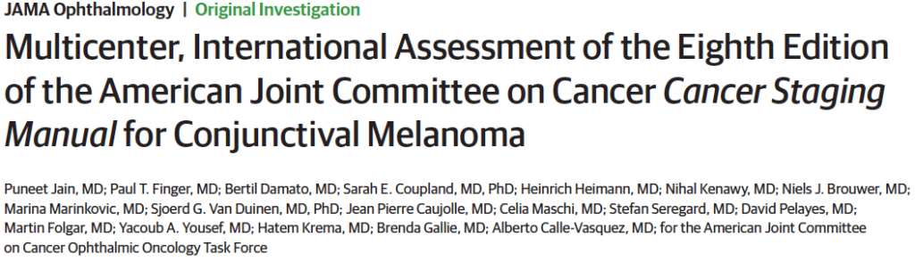
The American Joint Committee on Cancer (AJCC) Cancer Staging Manual compiles all currently available knowledge on cancer staging at various anatomic sites. In 2016, they released the 8th edition, conjunctival melanoma staging system which features 12 new staging systems, a wide range of new staging definitions, and an emphasis on the personalized-medicine approach. This [..] Read More...
Pakistan’s First Trained Ocular Oncologist
Pakistan is another country claimed by the Eye Cancer Foundation! Recently, Dr. Saima Amin graduated from her training sponsored by the Eye Cancer Foundation in Amman, Jordan at the King Hussain Cancer Center under the training of Dr. Yacoub Yousef. Of the hospital she worked, she notes that unfortunately in order to save their lives, [..] Read More...
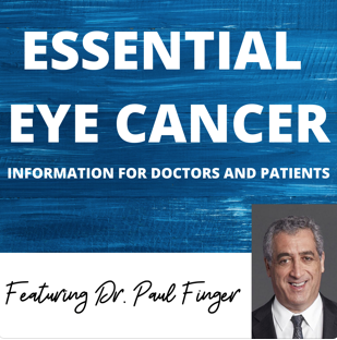
Podcasts from Dr. Finger!
Announcing the Essential Eye Cancer Podcast Page with Dr. Paul Finger! Dr. Finger has been working hard to give you this latest addition to the New York Eye Cancer website. For those of you who are auditory learners, these new Podcasts are where you can find your questions answered regarding Dr. Finger’s treatments, techniques and [..] Read More...
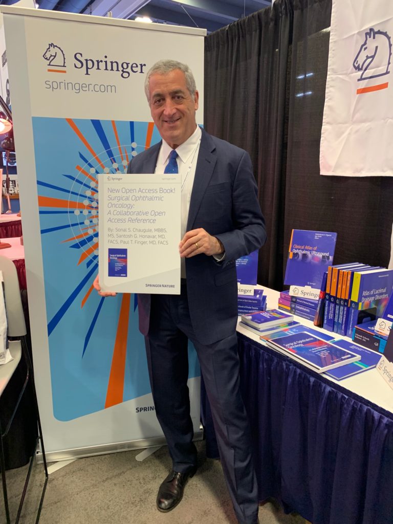
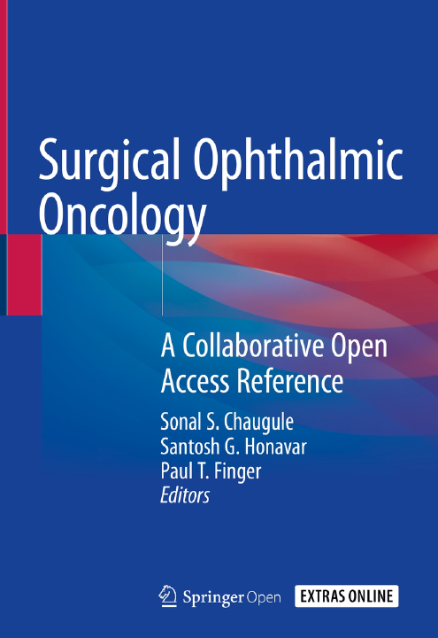
Surgical Ophthalmic Oncology: A Collaborative Open Access Reference is Published!
Surgical Ophthalmic Oncology: A Collaborative Open Access Reference has been published by Springer Nature and is available for download for free here! Five years ago, The First Eye Cancer Working Day was hosted by The Curie Institute in Paris, France. This was where the idea for the textbook was born, then called a “Collaborative Open [..] Read More...

“Best” Ophthalmologists in New York
Dr. Paul Finger of the New York Eye Cancer Center is consistently rated among the best doctors in the greater New York City region. As of 2023, Dr. Finger has appeared on the Castle Connolly list of “Top Doctors” in the New York area for the 16th time since 2005. Castle Connolly Medical Ltd., a [..] Read More...
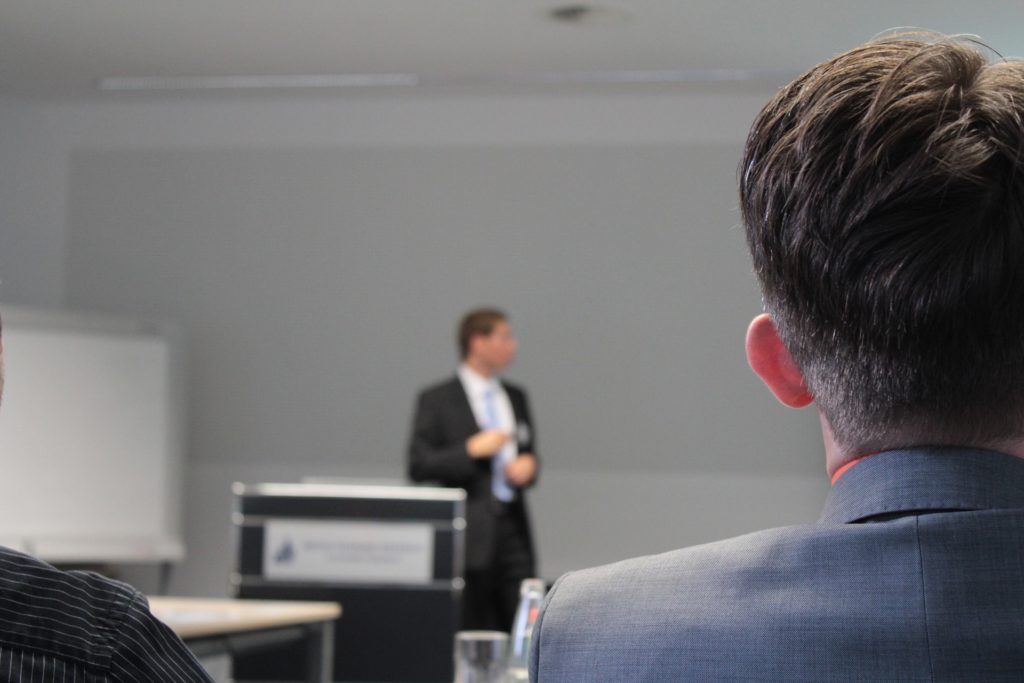
Annual Ophthalmic Oncology Lectureship
At the 41st Annual Tulane Eye Alumni Day at the Renaissance New Orleans Pere Marquette Hotel, Dr. Finger lectured about how and why his important innovations came to life. In the lecture, he discusses his early work on plaque microwave hyperthermia and TRT for intraocular tumors, pd-103 plaque radiation therapy, the slotted plaque, the amniotic [..] Read More...
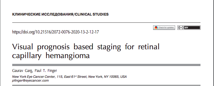
The Garg-Finger Staging System for Retina Capillary Hemangioma
Drs. Garg and Finger created the first visual acuity prognosis based staging system for retina capillary hemangioma (CRH). It appeared in the May 2020 issue of the Russian Ophthalmological Journal. Their study titled, “Visual Prognosis Based Staging for Retinal Capillary Hemangioma,” was derived from an analysis of the medical literature on Medline and PubMed. Thus, [..] Read More...
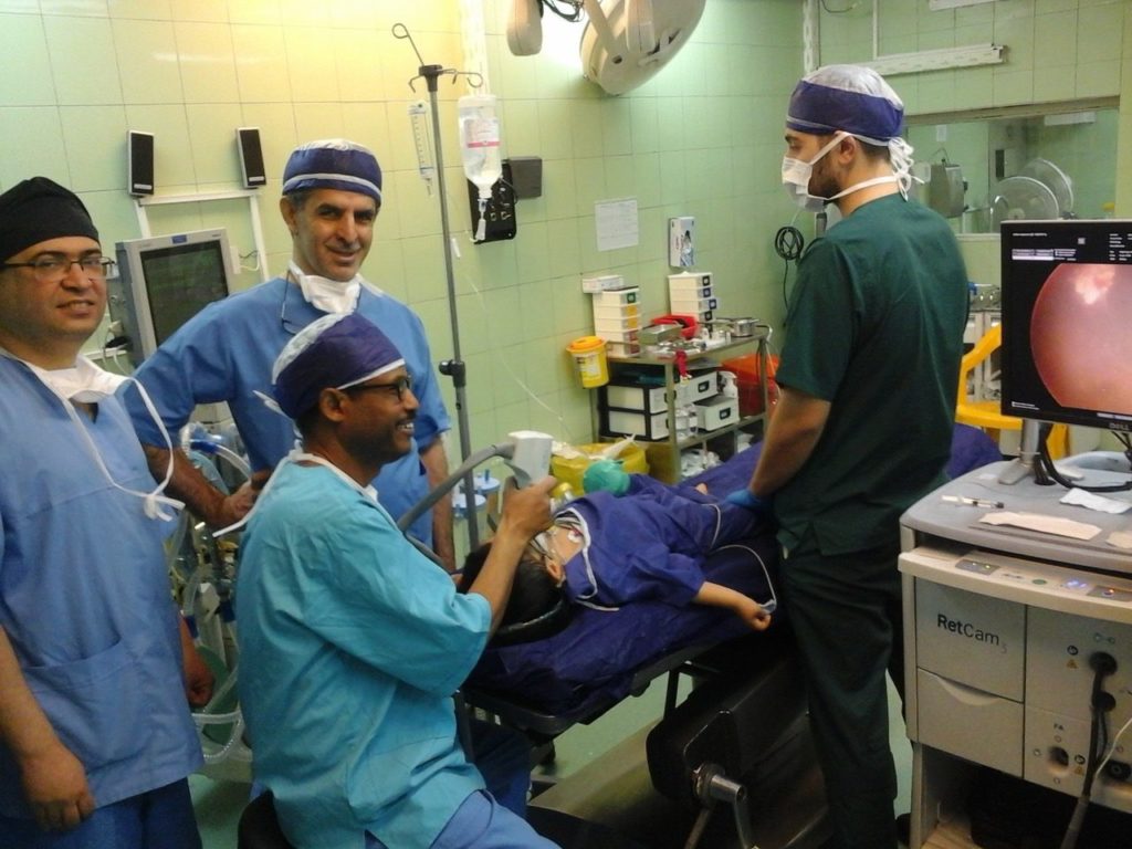
Eye Cancer Care in Ethiopia
Dr. Finger's Desk ECF Achievements
The Eye Cancer Foundation’s “2020 Campaign” claims another country, Ethiopia! We supported intensive training of a local ophthalmologist on advancements in retinoblastoma research, treatment, and diagnosis. Our most recent ECF grant recipient, Dr. Abu Amare, an ophthalmologist in Ethiopia, just completed a 6-month retinoblastoma fellowship at the Rasoole-E-Akram Hospital of The Iranian University of Medical [..] Read More...
Dr. Finger's Interview with The American Society of Retina Specialists
Barton Blackorby: You are the founding member of The New York Eye Cancer Foundation. Could you give some tips on how others can get involved in things like that and how you manage such a busy practice? Paul T. Finger: Back in 1998, I found myself leaving academic-based medicine and going into private practice. And [..] Read More...

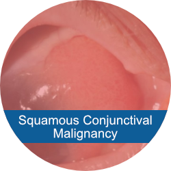
A "Biomarker" for Conjunctival Melanoma
The COMS Study What causes conjunctival melanoma (CoM)? Because of its rarity, much about CoM is unknown. Current medicine has yet to truly pinpoint any underlying genetic factors affecting CoM. In fact, no molecular drivers have been clearly defined in association with metastasis, recurrence prognosis, cell type, or other characteristic factors of CoM. In response [..] Read More...
Associate Specialist Opening at the NYECC
Join Dr. Finger and his team at the New York Eye Cancer Center (NYECC)! Currently, there is an opening for a fellowship trained associate specialist to join Dr. Finger in the practice of Ocular Tumor, Orbital Disease, and Ophthalmic Radiation Therapy. The ideal candidate should be able to treat patients with ocular tumors of the [..] Read More...

Eye Cancer Cluster in Raleigh, North Carolina
A recent article was just published in an online magazine called “Southerly” about a possible correlation between cancer incidence in Huntersville, North Carolina and industrial pollution. This relationship was scrutinized upon learning that several high school kids in a school by Lake Norman were diagnosed with ocular cancer. This prompted the residents to look for [..] Read More...
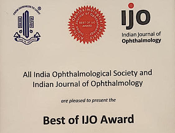
ECF Research wins Best of IJO Award!
In January of 2018, ECF fellow Dr. Sonal Chaugule, alongside Dr. Paul Finger and Dr. J. Park, published the study “Topical Chemotherapy for Giant Ocular Surface Squamous Neoplasia (OSSN) of the Conjunctiva and Cornea: Is Surgery Really Necessary?” in the Indian Journal of Ophthalmology (IJO). We are pleased to announce that this research has recently [..] Read More...
ECF Supported Research Featured in New York Eye and Ear Infirmary 200 Years Celebration
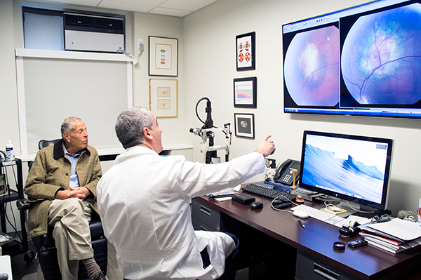
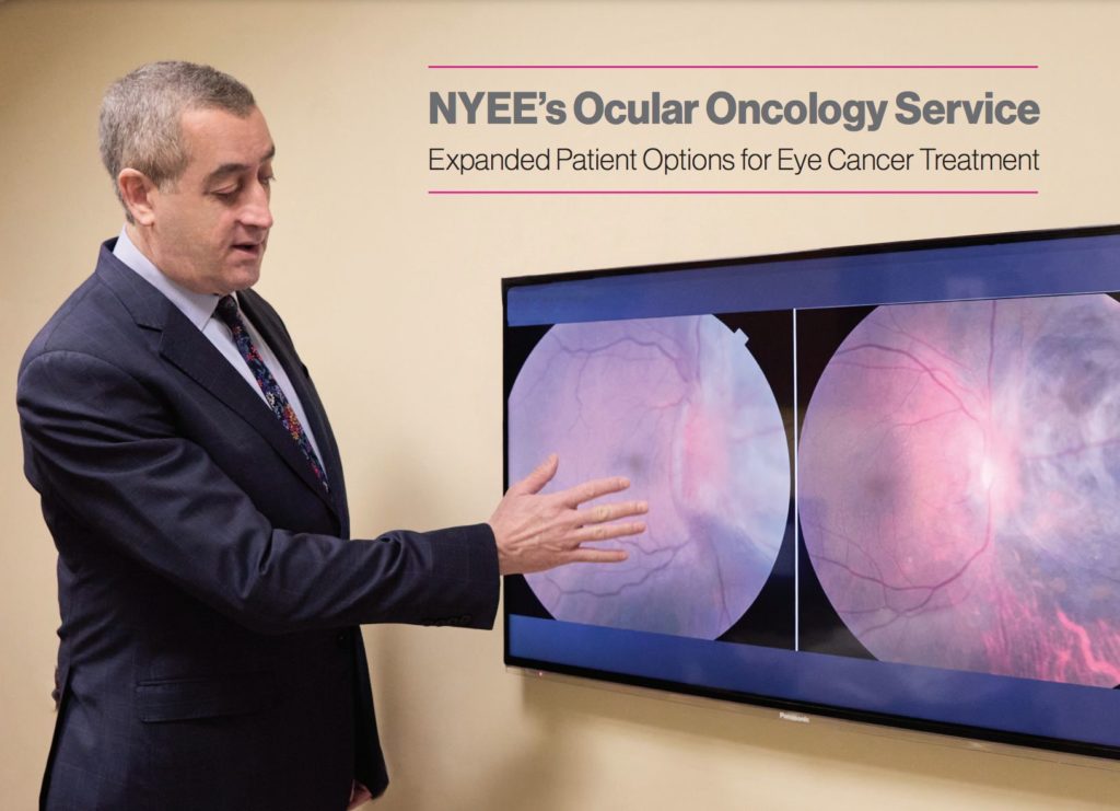
The New York Eye and Ear Infirmary of Mount Sinai is celebrating 200 years of providing innovative, first-class care to patients with ocular diseases. At the core of the infirmary’s Ocular Oncology Service stands ECF Executive Director Paul T. Finger, MD, who doubles as the committee’s founding director. The Eye Cancer Foundation has supported much [..] Read More...
RESULTS
Dr. Finger's Reported Outcomes
The following data represents near-real-time averages from all consecutive patients as they return to Dr. Finger’s office for follow up examinations (Last updated on ). About the data and disclaimer
CONDITIONS & TREATMENTS
Comprehensive Eye Cancer Information Covering eye cancer and related eye diseases—including symptoms, diagnosis, treatments, and much more. Search Conditions and TreatmentsMost Viewed Conditions
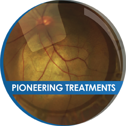
Treatment Types
- Enucleation Surgery - Removal of the Eye
- Ocular Prostheses Can Offer an Excellent Cosmetic Result
- Eye and Vision Sparing Radiation Therapy for Intraocular Tumors
- Intravitreal Anti-VEGF Therapy for Radiation Retinopathy
- Radiation Retinopathy Prevention and Suppression
- Proton Beam Versus Plaque
- Proton Beam Radiotherapy
- Systemic Chemotherapy
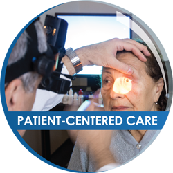
About Dr. Finger
Dr. Finger is an internationally recognized eye cancer specialist. His 35 years in ophthalmic oncology have been dedicated to learning, improving and inventing new methods for the diagnosis and treatment of cancers in and around the eye. These cancers are all rare and are commonly diagnosed without biopsy. Dr. Finger has spent his entire career caring for eye cancer patients. He has written hundreds of scientific publications and obtained patents for his original work.
OUR APPROACH

CONNECT AND SHARE


